SEM-Quanta , SEM-Fera , TEM-Tecnai , TEM-Jeol – are microscopes which is available in the LEM.
SEM-Quanta、SEM-Fera、TEM-Tecnai、TEM-Jeol – 是 文中提到的显微镜。
ACOM – Automated Crystal Orientation Mapping (TEM-Tecnai)
ACOM – 自动晶体取向映射 (TEM-Tecnai)
ACOM is based on electron diffraction using a quasi-parallel nanobeam to scan a selected region of interest. It is used for nanometer resolution mapping of (1) phase distribution in the sample (combined with EDS to determine chemical composition), (2) crystallographic orientation, and (3) stress distribution in a single-phase sample.
ACOM 基于电子衍射,使用准平行纳米束扫描选定的感兴趣区域。它用于对 (1) 样品中的相分布(与 EDS 结合以确定化学成分)、(2) 晶体取向和 (3) 单相样品中的应力分布进行纳米分辨率绘图。
BF / DF – Bright-Field / Dark-Field Imaging (TEM-Tecnai, TEM-Jeol)
BF / DF – 明场/暗场成像(TEM-Tecnai、TEM-Jeol)
BF/DF is a basic method of imaging crystalline materials in TEM based on diffraction contrast. By using an objective aperture, which selects only one electron beam for imaging, it strongly enhances the image contrast (BF). Using directed sample tilting, precipitates in the matrix can be highlighted or dislocations/defects in the material structure can be studied.
BF/DF 是基于衍射对比度在 TEM 中对晶体材料进行成像的基本方法。通过使用仅选择一束电子束进行成像的物镜孔径,可以大大增强图像对比度 (BF)。使用定向样品倾斜,可以突出显示基体中的沉淀物,或者可以研究材料结构中的位错/缺陷。
EBIC – Electron Beam Induced Current (SEM-Fera)
EBIC – 电子束感应电流 (SEM-Fera)
EBIC is a method in which an electron beam induces a current in the sample that can be used as a signal to create images describing the electrical characteristics of the sample, e.g. areas of PN transitions in the sample, presence of local defects, contaminations, inhomogeneity of impurities, or to investigate properties of minority charge carriers.
EBIC 是一种电子束在样品中感应电流的方法,该电流可用作信号来创建描述样品电气特性的图像,例如样品中 PN 跃迁的区域、局部缺陷的存在、污染、杂质的不均匀性,或研究少数电荷载流子的特性。
EBSD – Electron Back-Scatter Diffraction (SEM-Quanta, SEM-Fera)
EBSD – 电子背散射衍射(SEM-Quanta、SEM-Fera)
EBSD uses diffraction of back-scattered electrons in the form of Kikuchi lines to obtain information about the structure of the sample. It is used in SEM to study phase composition, crystallographic orientation or stress in materials. For example, the phase composition and preferred orientation of crystals in 3D-printed samples of metallic materials can be studied.
EBSD 利用菊池线形式的背散射电子衍射来获取有关样品结构的信息。它在 SEM 中用于研究材料中的相组成、晶体取向或应力。例如,可以研究 3D 打印金属材料样品中晶体的相组成和择优取向。
ED – Electron Diffraction (TEM-Tecnai, TEM-Jeol)
ED – 电子衍射(TEM-Tecnai、TEM-Jeol)
ED is used to differentiate phases with different atomic structures, e.g. graphite (C – hexagonal system) and diamond (C – cubic system). Three basic types of electron diffraction are distinguished: (1) spot diffraction patterns for single crystal samples, (2) ring diffraction patterns for powder samples, and (3) diffraction halo for amorphous samples. To determine the unknown structure, a 3D ED technique is used in which the sample is sequentially tilted while the ED patterns are recorded.
ED 用于区分具有不同原子结构的相,例如石墨(C – 六方晶系)和金刚石(C – 立方晶系)。电子衍射分为三种基本类型:(1) 单晶样品的点衍射图案,(2) 粉末样品的环形衍射图案,以及 (3) 非晶样品的衍射晕。为了确定未知结构,使用了 3D ED 技术,在记录 ED 图案的同时顺序倾斜样品。
EDX ( EDS ) – Energy Dispersive X-ray Spectroscopy (SEM-Quanta, SEM-Fera, TEM-Tecnai)
EDX ( EDS ) – 能量色散 X 射线光谱(SEM-Quanta、SEM-Fera、TEM-Tecnai)
EDS is a method for determining the chemical composition of a sample that is used in both SEM and S/TEM. It allows for the detection of elements from B upwards with a detection limit of 0.x-0.0x wt% from regions of about a micrometre (in the case of SEM) and about a nanometer (in the case of S/TEM). Elemental maps can be generated in SEM and STEM, and in combination with SEM/FIB, the surface of the sample can be sequentially removed by ions to obtain 3D elemental maps.
EDS 是一种用于确定样品化学成分的方法,可用于 SEM 和 S/TEM。它允许从大约一微米(在 SEM 的情况下)和大约一纳米(在 S/TEM 的情况下)区域检测 B 向上的元素,检测限为 0.x-0.0x wt%。可以在SEM和STEM中生成元素图,并与SEM/FIB结合,可以通过离子顺序去除样品表面以获得3D元素图。
EELS – Electron Energy Loss Spectrometry (TEM-Tecnai)
EELS – 电子能量损失光谱法 (TEM-Tecnai)
EELS is a method for determining the chemical composition of a sample that is used in S/TEM. It complements and extends the information obtained by EDS. It allows for the detection of light elements (H, He, Li) and is particularly useful in case of overlapping signals in EDS (e.g. Ti and N). Furthermore, it is possible to distinguish the same element in different structural modifications or with different valence. Elemental maps can also be created in STEM.
EELS 是一种确定 S/TEM 中使用的样品化学成分的方法。它补充并扩展了 EDS 获得的信息。它可以检测轻元素(H、He、Li),并且在 EDS 中信号重叠(例如 Ti 和 N)的情况下特别有用。此外,可以区分不同结构修饰或不同化合价的相同元素。元素图也可以在 STEM 中创建。
EFTEM – Energy Filtered Transmission Electron Microscopy (TEM-Tecnai)
EFTEM – 能量过滤透射电子显微镜 (TEM-Tecnai)
EFTEM is used to display the distribution of elements in the sample. It is based on an EEL spectrum from which a selected signal (energy region) can be filtered out using a slit. In EFTEM, a broad parallel beam is used and the image is formed instantaneously, unlike in STEM/EELS where a convergent beam is used and the region of interest is scanned sequentially. However, at higher electron energy losses, the signal is often too weak for EFTEM.
EFTEM 用于显示样品中元素的分布。它基于 EEL 光谱,可以使用狭缝从中滤除选定的信号(能量区域)。在 EFTEM 中,使用宽的平行光束并立即形成图像,这与 STEM/EELS 不同,在 STEM/EELS 中使用会聚光束并顺序扫描感兴趣区域。然而,在电子能量损失较高的情况下,对于 EFTEM 来说信号通常太弱。
FIB – Focused Ion Beam (SEM-Quanta, SEM-Fera)
FIB – 聚焦离子束(SEM-Quanta、SEM-Fera)
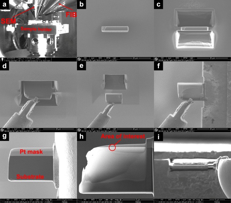
FIB is very often used in combination with SEM (however, there are also single-beam instruments available using only FIB). FIB is used for: (1) sequential removal of sample surface in 3D methods (imaging, EBSD, EDS, EBIC), (2) creating cross-sections, (3) nano-machining – e.g. preparation of nanopillars, and (4) preparation of TEM lamellas. There are two types of FIB: (1) Liquid Metal Ion Source (LMIS) – more suitable for fine work, and (2) Plasma – more suitable for the removal of large volumes while reducing the contamination.
FIB 通常与 SEM 结合使用(但是,也有仅使用 FIB 的单光束仪器)。 FIB 用于:(1) 以 3D 方法(成像、EBSD、EDS、EBIC)顺序去除样品表面,(2) 创建横截面,(3) 纳米加工 – 例如纳米柱的制备,以及(4)TEM 片层的制备。 FIB 有两种类型:(1) 液态金属离子源 (LMIS) – 更适合精细工作,(2) 等离子体 – 更适合去除大量样品,同时减少污染。
HAADF – High Angle Annular Dark Field (TEM-Tecnai)
HAADF – 高角度环形暗场 (TEM-Tecnai)
HAADF is used in STEM mode to detect electrons scattered to high angles. This is incoherent scattering on the atoms of the sample, which shows a dependence on the square of the atomic number. Therefore, elements with higher atomic number scatter at higher angles. Thus, it is easy to distinguish the parts of the sample with light elements from the parts with heavy elements, e.g. Pd catalyst particles on Al2O3 support. In state-of-the-art microscopes, it is possible to focus the beam below the size of an atom and therefore “map” individual atoms directly.
HAADF 在 STEM 模式下用于检测散射到大角度的电子。这是样品原子上的不相干散射,显示出对原子序数平方的依赖性。因此,原子序数较高的元素以较高的角度散射。因此,很容易区分样品中含有轻元素的部分和含有重元素的部分,例如Al2O3 载体上的 Pd 催化剂颗粒。在最先进的显微镜中,可以将光束聚焦到原子大小以下,从而直接“绘制”单个原子。
HRTEM – High Resolution Transmission Electron Microscopy (TEM-Tecnai)
HRTEM – 高分辨率透射电子显微镜 (TEM-Tecnai)
HRTEM is an imaging method in the TEM using a wide parallel beam. The image is formed by interference of multiple diffracted beams. When the crystal sample is properly aligned (the atoms are aligned in columns parallel to the incident electron beam), the result is a periodic image with atomic resolution. The HRTEM has applications, for exmaple, in the study of dislocations/defects in the structure, imaging of twin lamellae or the study of thin films.
HRTEM 是一种使用宽平行光束的 TEM 成像方法。图像是由多个衍射光束干涉形成的。当晶体样品正确排列时(原子排列成平行于入射电子束的列),结果是具有原子分辨率的周期性图像。 HRTEM 可应用于结构中的位错/缺陷研究、双片层成像或薄膜研究。
LTEM – Lorentz Transmission Electron Microscopy (TEM-Tecnai)
LTEM – 洛伦兹透射电子显微镜 (TEM-Tecnai)
LTEM is used to observe magnetic domain structures. The trajectories of electrons flying through the sample region with magnetic induction are deflected by the Lorentz force. To visualize the domains, we mostly use the defocusing (Fresnel) method. Thus, if we focus on a plane close below or above the sample, light and dark fringes corresponding to the boundaries of the individual domains appear in the image due to the phase shift of the individual rays.
LTEM 用于观察磁畴结构。电子在磁感应作用下飞过样品区域的轨迹会因洛伦兹力而发生偏转。为了可视化域,我们主要使用散焦(菲涅耳)方法。因此,如果我们聚焦在靠近样本下方或上方的平面上,由于各个光线的相移,与各个域的边界相对应的明暗条纹会出现在图像中。
SEM – Scanning Electron Microscopy (SEM-Quanta, SEM-Fera)
SEM – 扫描电子显微镜(SEM-Quanta、SEM-Fera)
SEM studies the surface of samples by scanning a focused electron beam. Electrons interact with the sample and a large number of types of signal are produced. The basic types of imaging are (1) in secondary electrons (SE), which are sensitive to the surface morphology, and (2) in backscattered electrons (BSE), which show contrast due to the average atomic number. In addition, techniques for determining chemical composition (EDS) and detecting crystallographic orientation (EBSD) are used in SEM. In materials science, coupling with FIB is often used due to extending the 2D analysis into 3D by sequentially removing the sample surface.
SEM 通过扫描聚焦电子束来研究样品的表面。电子与样品相互作用并产生多种类型的信号。成像的基本类型是(1)二次电子(SE),对表面形态敏感,以及(2)背散射电子(BSE),由于平均原子序数而显示对比度。此外,SEM 中还使用了确定化学成分 (EDS) 和检测晶体取向 (EBSD) 的技术。在材料科学中,由于通过顺序去除样品表面将 2D 分析扩展到 3D,因此经常使用与 FIB 的耦合。
STEM – Scanning Transmission Electron Microscopy (TEM-Tecnai)
STEM – 扫描透射电子显微镜 (TEM-Tecnai)
The image in STEM is created by scanning a focused beam over a thin sample (transparent to electrons) and detecting the transmitted electrons (BF, ADF, HAADF). In state-of-the-art microscopes, it is possible to focus the beam below the size of an atom and therefore directly “map” individual atoms. In our lab, STEM is mainly used for chemical composition mapping by EDS and EELS and imaging by HAADF.
STEM 中的图像是通过在薄样品(对电子透明)上扫描聚焦光束并检测透射电子(BF、ADF、HAADF)来创建的。在最先进的显微镜中,可以将光束聚焦到原子大小以下,从而直接“绘制”单个原子。在我们的实验室中,STEM 主要用于 EDS 和 EELS 化学成分绘图以及 HAADF 成像。
TEM – Transmission Electron Microscopy (TEM-Tecnai, TEM-Jeol)
TEM – 透射电子显微镜(TEM-Tecnai、TEM-Jeol)
TEM studies thin samples with the electron beam that passes through the sample. The electrons interact with the sample and a large number of types of signals are produced, which allow for the study of not only the microstructure of samples down to atomic resolution (BF, DF, HRTEM), but also the chemical composition (EDS, EELS) or the crystallographic orientation of individual grains (ED, ACOM).
TEM 使用穿过样品的电子束来研究薄样品。电子与样品相互作用并产生大量类型的信号,这不仅可以研究样品的微观结构,达到原子分辨率(BF、DF、HRTEM),还可以研究化学成分(EDS、EELS) )或单个晶粒的晶体取向(ED、ACOM)。

